Fields covered: Neuroscience, Neuroanatomy, Neuronal Signalling, Neuronal Development
Related Articles:
§ How to map the brain §
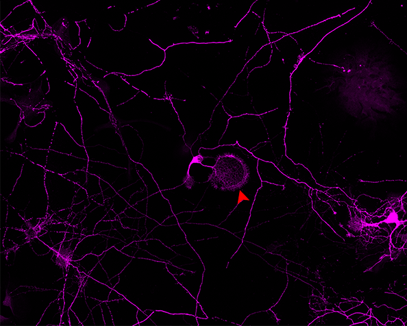
![]() Growth Cones §
Growth Cones § ![]() Cytoplasmic mechanisms of axonal and dendritic growth in neurons §
Cytoplasmic mechanisms of axonal and dendritic growth in neurons § ![]() Cytoskeletal events in growth cone steering|
Cytoskeletal events in growth cone steering|